Introduction: Understanding Gastroshiza in Modern Medicine
Gastroshiza, more commonly and medically referred to as gastroschisis, is a congenital condition that has drawn the attention of clinicians, researchers, and expecting families worldwide due to its complex clinical presentation and the challenges involved in early diagnosis and surgical management. At its core, gastroshiza describes a situation in which an infant’s abdominal wall fails to develop completely during fetal life, creating an opening through which the intestines and sometimes other visceral organs protrude outside the body without any protective covering. This defect typically occurs to the right of the belly button and is distinguishable from other ventral wall defects such as omphalocele, in which the exposed organs are contained within a membrane. The absence of a protective sac in gastroshiza exposes the intestines to amniotic fluid during pregnancy, which often leads to inflammation, irritation, and varying degrees of organ dysfunction even before birth. While the exact cause of gastroshiza remains unclear, research suggests that a combination of genetic, environmental, and placental factors may play roles in its development, with incidence estimates ranging from approximately one in every 2,500 to 5,000 live births in populations studied worldwide. Early prenatal detection using advanced ultrasonography has become a cornerstone of modern obstetric care, allowing healthcare teams to prepare comprehensive delivery and surgical plans. Today, thanks to advances in neonatal surgery and multidisciplinary neonatal intensive care, survival rates for infants born with gastroshiza have improved significantly. Nevertheless, the condition continues to require careful management and long-term follow-ups to address potential complications such as feeding difficulties, growth concerns, and gastrointestinal dysfunction, making it both a medical and psychosocial focus at the intersection of perinatal medicine and pediatric surgery.
The Clinical Definition and Pathophysiology of Gastroshiza
At its most fundamental level, gastroshiza is defined as a full-thickness paraumbilical abdominal wall defect through which abdominal organs protrude, most frequently small intestine loops, but occasionally including stomach and liver segments, into the perinatal environment outside the fetus’s body. This defect arises early in gestation, typically during the first trimester, when the embryonic lateral body wall folds fail to fuse correctly at the midline, leaving a persistent opening adjacent to the umbilicus. While the precise molecular and embryologic triggers for this failure of closure remain under investigation, contemporary studies indicate that the exposed bowel’s continuous contact with amniotic fluid results in progressive irritation, inflammation, and sometimes fibrosis, which can complicate both prenatal development and postnatal surgical repair. Unlike omphalocele, another congenital ventral wall defect, gastroshiza’s defining characteristic is the absence of a protective sac covering the extruding organs; this absence increases the risk of infection and fluid loss at birth and necessitates urgent clinical intervention. The lack of a sac likely reflects a direct effect of a primary abdominal wall closure defect rather than a failure of organ return from the umbilical cord. Biologically, the condition is associated with a spectrum of pathophysiologic changes, including chemical peritonitis and significant alterations in bowel structure and motility, which can have cascading effects on neonatal digestion and absorption. Modern embryologic theory also includes hypotheses about vascular disruption and abnormal mesodermal development, but no single cause has been definitively identified, underscoring the complexity of gastroshiza as a congenital anomaly whose roots lie in intricate early fetal development processes.
Epidemiology: Who Gets Gastroshiza and Why It Matters
In medical and public health analysis, understanding the epidemiology of gastroshiza is crucial not only for clinicians and surgeons but also for researchers studying congenital anomaly patterns worldwide. Broad epidemiologic data indicate that the incidence of gastroshiza varies slightly across geographic regions and population groups, but on average affects roughly one in 2,500 to one in 5,000 live births in high-income countries with sophisticated prenatal detection protocols. This rare yet consistently observed frequency situates gastroshiza among the more common abdominal wall defects encountered in neonatal medicine, but it remains a significant contributor to perinatal morbidity and intensive care utilization due to its need for immediate surgical correction after birth. Epidemiologic surveillance has revealed patterns that point to increased prevalence in babies born to younger mothers, particularly those under the age of twenty, and correlations with specific lifestyle factors such as maternal tobacco and alcohol use during pregnancy, although these associations do not yet clarify causation. Studies suggest that younger maternal age, low body mass index, and certain environmental exposures may influence the risk of developing gastroshiza, but the underlying mechanisms remain subjects of ongoing research. Additionally, the prevalence of gastroshiza has appeared to rise slowly over recent decades in some countries, prompting public health researchers to explore links with broader socioeconomic, behavioral, and environmental trends. While the condition itself is generally considered isolated and not commonly associated with genetic syndromes, there are instances where additional congenital anomalies such as intestinal atresia or malrotation may accompany gastroshiza, and these cases often incur greater clinical complexity and longer hospital stays. The epidemiologic profile of gastroshiza, therefore, underscores not just its medical significance but also its relevance to maternal health education, prenatal care planning, and long-term pediatric outcomes.
Etiology: Exploring the Causes Behind Gastroshiza
The etiology of gastroshiza remains one of the most intriguing aspects of this congenital condition, blending elements of embryology, genetics, and environmental science in ongoing research efforts seeking to uncover its origins. Fundamentally, gastroshiza arises due to incomplete development of the ventral abdominal wall during the early weeks of embryogenesis, but beyond this anatomical description lies a web of unanswered questions about why this failure occurs in some pregnancies and not others. Medical literature consistently reports that the exact cause of gastroshiza cannot yet be pinpointed to a single genetic mutation or environmental exposure, suggesting that a multifactorial process is most likely responsible. Large health studies underscore that a combination of genetic predispositions and environmental influences, such as maternal substance use, nutritional factors, and possibly infections, may interact to disrupt normal abdominal wall formation in utero. For example, maternal smoking and alcohol consumption during early pregnancy have been associated with increased risk, though these associations do not definitively show causality and may reflect broader lifestyle and socioeconomic conditions. Emerging data from genomic studies have begun to explore whether subtle gene regulatory differences might contribute to the defect, but to date, no consistent pathogenic variants have been identified as primary drivers in most cases. Some experts also theorize that disruptions in blood supply to the developing abdominal wall during critical periods of gestation may impede necessary tissue formation, setting the stage for gastroshiza. Despite these hypotheses, the medical community continues to emphasize that no specific cause can be universally applied, and each case may arise from a unique constellation of risk factors. As research grows more sophisticated, the enigma of gastroshiza’s etiology remains a central focus for both scientists and clinicians aiming to better understand and ultimately prevent this complex birth defect.
Prenatal Diagnosis: Detection Before Birth
Modern prenatal care places a significant emphasis on early identification of structural abnormalities, and gastroshiza is among the conditions most reliably detected before birth through routine imaging and maternal-fetal screening protocols. Typical prenatal diagnosis of gastroshiza occurs during second-trimester ultrasound examinations, often between the 18th and 22nd weeks of gestation, when detailed visualization of the fetus can reveal loops of intestine floating freely in amniotic fluid outside the fetal abdominal wall. This absence of a protective membrane and the distinct location of the defect — usually to the right of the belly button — make it possible for experienced sonographers to identify gastroshiza with a high degree of confidence. Prenatal diagnosis not only confirms the presence of the abdominal wall defect but also allows clinicians to assess the extent of organ exposure, potential complications such as intestinal edema or twisting, and the overall wellbeing of the fetus. In some cases, elevated maternal serum alpha-fetoprotein levels detected in routine blood screening can raise early suspicion of a ventral wall defect, prompting focused ultrasound investigations. When gastroshiza is diagnosed prenatally, it triggers a multidisciplinary care plan involving obstetricians, neonatologists, pediatric surgeons, and perinatal specialists to optimize delivery timing and location. Because infants with gastroshiza require immediate postnatal surgical repair and intensive care support, prenatal detection ensures that births take place in facilities equipped with specialized neonatal surgical and critical care services rather than settings lacking such resources. Additionally, prenatal counseling provides families with essential information about the condition, anticipated treatments, potential outcomes, and the comprehensive care pathway ahead, enabling informed decision-making. The ability to diagnose gastroshiza before birth represents a major advancement in perinatal medicine and reflects the broader progress in imaging technology and multidisciplinary obstetric care that has transformed outcomes for congenital anomalies in recent decades.
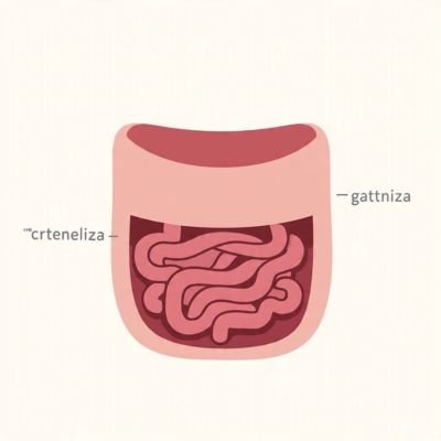
Clinical Presentation: Recognizing Gastroshiza at Birth
Infants born with gastroshiza exhibit a striking and immediately recognizable physical presentation, which often compels urgent clinical attention in delivery rooms. The hallmark feature is the protrusion of intestinal loops and, occasionally, segments of other abdominal organs through an opening typically located just to the right of the umbilicus. Unlike other ventral wall defects such as omphalocele, these organs are exposed directly to the external environment, without a protective membranous covering, making them highly susceptible to dehydration, trauma, and infection. The exposed bowel often appears swollen, thickened, and inflamed due to prolonged contact with amniotic fluid during gestation, which can trigger chemical irritation and peritoneal inflammation. In addition to the visible defect, affected neonates may exhibit complications related to gastrointestinal function, including delayed feeding tolerance, abnormal motility, or partial obstruction, which can become apparent within the first hours to days of life. Although gastroshiza is most often isolated, clinicians remain vigilant for accompanying anomalies, such as intestinal atresia, malrotation, or extraintestinal malformations, which can complicate both surgical management and long-term prognosis. Physical examination at birth is typically sufficient for initial diagnosis, but additional imaging may be employed to assess the integrity of the bowel, the presence of twisting or ischemia, and other structural concerns. Prompt recognition of gastroshiza is critical, as it sets the stage for immediate stabilization, including covering the exposed intestines with sterile, moist dressings, monitoring fluid and electrolyte balance, and planning surgical intervention. In modern neonatal care, this early recognition, combined with a multidisciplinary approach, has dramatically improved both survival and quality-of-life outcomes for infants affected by gastroshiza.
Surgical Management: Techniques and Considerations
Surgical intervention for gastroshiza is universally recognized as the cornerstone of treatment, with the primary goal being the safe and complete return of extruded abdominal contents into the peritoneal cavity followed by closure of the abdominal wall defect. The timing, technique, and complexity of surgery depend on factors such as the size of the defect, the number of organs involved, the degree of bowel inflammation, and the overall condition of the neonate. In some cases, surgeons employ a one-stage primary closure, in which the intestines are carefully repositioned, and the abdominal wall is closed immediately after birth. In other scenarios, a staged approach, often involving the use of a protective silo, is preferred, allowing gradual reduction of the intestines into the abdominal cavity over several days to prevent abdominal compartment syndrome and reduce tension on the tissues. Meticulous care is taken to preserve bowel integrity, minimize trauma, and prevent postoperative complications such as adhesions, necrosis, or infection. Postoperative care in a neonatal intensive care unit is critical, involving close monitoring of fluid and electrolyte balance, pain management, and gradual introduction of enteral nutrition once bowel function resumes. Advances in surgical techniques, including minimally invasive approaches and innovative closure materials, have further refined outcomes, reducing complications and hospitalization times. Collaboration between surgeons, anesthesiologists, nurses, and pediatric specialists ensures that each infant’s care plan is individualized, taking into account both immediate surgical needs and long-term gastrointestinal function. Surgical management not only restores anatomical integrity but also serves as the foundation for optimizing long-term growth, nutrition, and overall health, highlighting the importance of timely and expert intervention in the treatment of gastroshiza.
Postoperative Care and Long-Term Management
The journey of an infant with gastroshiza does not conclude with surgical repair; rather, postoperative care and long-term management play a critical role in determining outcomes and quality of life. Following surgery, neonates are closely monitored in specialized intensive care settings to manage fluid balance, infection prevention, and pain control. The exposed bowel may initially be slow to regain full motility, necessitating careful management of nutrition through parenteral feeding until the gastrointestinal tract is capable of handling enteral feeding. Long-term follow-up often involves monitoring growth parameters, bowel function, and developmental milestones, as some infants may experience complications such as short bowel syndrome, malabsorption, or feeding intolerance. Families are educated regarding signs of infection, dehydration, and bowel obstruction, ensuring early intervention if problems arise. Psychological support and counseling for parents and caregivers are also integral, as the stress associated with prolonged hospitalization, complex surgeries, and ongoing care can be significant. In addition, coordinated care among pediatricians, surgeons, nutritionists, and gastroenterologists provides a framework for addressing any late-emerging concerns, optimizing developmental outcomes, and supporting the transition from hospital to home. Continuous research and clinical audits help refine management protocols, identify risk factors for complications, and implement best practices that enhance survival rates and long-term well-being. In sum, effective postoperative care and vigilant long-term management are essential in transforming gastroshiza from a high-risk neonatal condition into a manageable medical challenge, with the potential for affected children to lead healthy, thriving lives.
Prognosis and Outcomes in Contemporary Medicine
Historically, gastroshiza was associated with high mortality rates due to sepsis, dehydration, and complications arising from exposed abdominal organs, but contemporary medicine has transformed its prognosis remarkably. Advances in prenatal detection, surgical techniques, and neonatal intensive care have collectively increased survival rates to over 90% in high-resource settings. The outcome for each infant is influenced by factors such as the size of the defect, condition of the protruding organs, presence of additional anomalies, and timeliness of medical intervention. Infants who undergo successful surgical repair and receive comprehensive postoperative care often experience normal growth and development, although some may require ongoing nutritional support or monitoring for gastrointestinal motility issues. Longitudinal studies suggest that most children with isolated gastroshiza enjoy quality of life comparable to their peers, with minimal limitations in physical activity or daily function. Nonetheless, ongoing research emphasizes the importance of optimizing early interventions, improving surgical techniques, and providing multidisciplinary support to address potential complications, particularly in settings with limited medical resources. The evolution of outcomes over the past decades highlights both the resilience of affected infants and the profound impact of coordinated medical care. For parents and clinicians alike, the modern prognosis for gastroshiza underscores the value of early diagnosis, prompt surgical repair, meticulous postoperative care, and a holistic approach to pediatric health, demonstrating that a condition once fraught with risk can now be managed effectively with proper expertise and infrastructure.
Conclusion: Awareness, Research, and the Path Forward
Gastroshiza represents a complex, multifactorial congenital anomaly that challenges healthcare systems, families, and researchers alike, yet advances in prenatal detection, surgical intervention, and neonatal care have redefined its trajectory and improved outcomes substantially. The condition’s early recognition, both prenatally and immediately after birth, is critical for minimizing complications and ensuring successful intervention, while a multidisciplinary care model encompassing surgeons, neonatologists, nurses, and family support structures has become the standard for effective management. Ongoing research into the underlying causes, whether genetic, environmental, or vascular, continues to inform prevention strategies, clinical protocols, and counseling for affected families. Awareness campaigns, evidence-based clinical guidelines, and the integration of modern imaging and surgical techniques have collectively transformed gastroshiza from a high-risk, life-threatening condition into one that is manageable with appropriate expertise and timely care. The journey from understanding etiology, providing skilled surgical repair, to delivering long-term support highlights the remarkable progress achieved in perinatal medicine. At Infoaxis, we are committed to providing authoritative information, educating families and healthcare providers, and supporting research into conditions like gastroshiza to ensure informed decision-making, improved care standards, and better outcomes for affected infants and their families. By fostering awareness and advancing knowledge, we contribute to the broader mission of transforming congenital conditions into manageable medical challenges through collaboration, innovation, and compassionate care.


Leave a Reply